
When Conventional X-Rays May Miss the MarkMatt Kroona, X.T.
Orange Coast Dental X-ray Lab Huntington Beach CA From the Summer 2009 AADMRT Newsletter  Matt Kroona Matt Kroona
While accompanying his sister to a routine orthodontic appointment, this 16 year old male patient casually mentioned to the orthodontist that he had a "bump" in his gums on the side of his mouth, above tooth #3. Following an examination, the patient was referred to an imaging center for occlusal, periapical, and bitewing x-rays. (figs. 1 & 2).
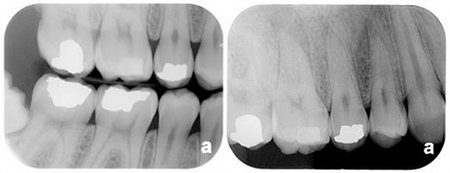
Figure 1
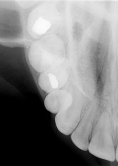
Figure 2
|


|
After reviewing the conventional radiographs, the orthodontist prescribed a Cone Beam CT scan of the maxilla. Upon completion of the procedure, it became clear what the conventional x-rays had failed to completely reveal.
The reconstructed panoramic view (fig. 3) shows possible problems in the apical region of tooth #3 as well as possible involvement in the sinus.
The reconstructed panoramic view (fig. 3) shows possible problems in the apical region of tooth #3 as well as possible involvement in the sinus.
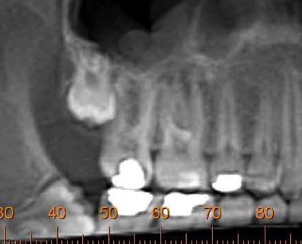
Figure 3
The cross sections (fig. 4) clearly reveal apical lesions in the distobuccal root (#61), the lingual root (#63 & #65), and the mesiobuccal root (#67). Also seen in the cross sections is the fact that the floor of the right maxillary sinus is discontinuous, and that the lucency at the apices is continuous with the lumen (air space) of the sinus. There is also a soft tissue density in the floor of the sinus.
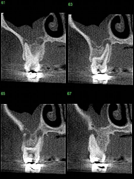
Figure 4
The 3D rendering that was also provided (fig. 5) helped the patient better understand the "bump" he was feeling and what was happening in his mouth.
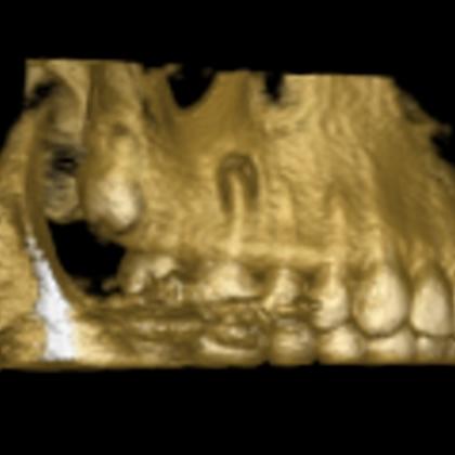
Figure 5
With the help of the CBCT images, this patient was referred (along with the images) to an endodontist for treatment.
This case demonstrates that when limited to conventional dental x-rays, the apparent "absence of evidence" (in this case pathology) is not necessarily the "evidence of absence." Through the combination of an observant orthodontist and the availability of modern imaging technology, a possible serious problem may have been avoided.
This case demonstrates that when limited to conventional dental x-rays, the apparent "absence of evidence" (in this case pathology) is not necessarily the "evidence of absence." Through the combination of an observant orthodontist and the availability of modern imaging technology, a possible serious problem may have been avoided.