
From Japan: Newly added oral
|


|
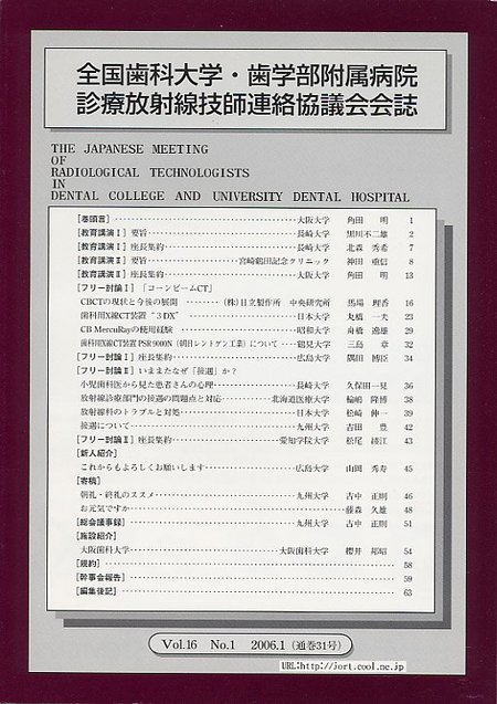
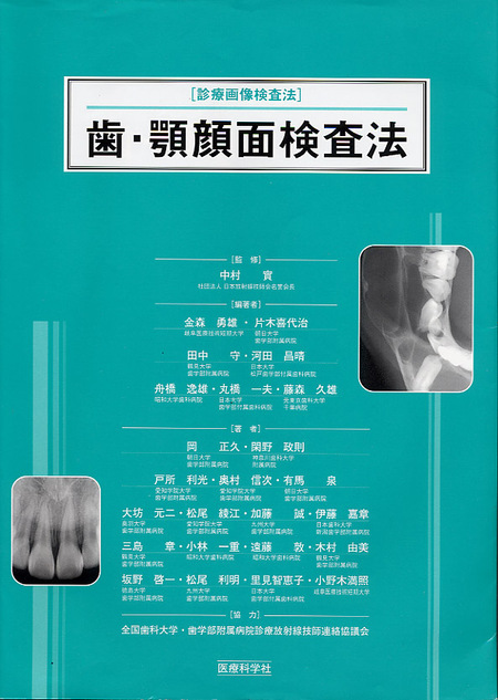
The Publisher's website is:
http://www.iryokagaku.co.jp/frame/03-honwosagasu/shuyoumokuji-860033078/shuyoumokuji-860033078.html
The website is written in Japanese, but the images will perhaps be of interest even if you cannot understand the text.
New CBCT System (1): "Alphard"
The number of cone-beam CT units installed in dental offices in Japan is seemed to be between 200 and 300 now. J. Morita and Hitachi Medical Systems are no the only manufacturers of dental cone-beam CT systems in Japan, Asahi Roentgen Industry is also a manufacturer having unique concepts. The sales company in the United States is Belmont.
The Asahi's first CBCT unit, PSR9000N, was released several years ago. The website is http://www.asahi-xray.co.jp/global/psr9000n.htm. To my knowledge, technical assessments and clinical studies have never been published in academic journals written in English. But Asahi is a well-known company in Japan.
The outstanding characteristic is thought to be what is described as the "panoramic CT function." This is achieved with a continuous 7-times 360-degree rotation, while the rotation center is gradually shifting, producing a cross-sectional tubular area of 34x40mm along the full dento-alveolar arch. The total exposure time is only 30 seconds for this 7-times 360-degree rotation. Subsequently, seven "Dental CT" images having the dimension, 40(height)x41 mm (in diameter), are continuously set out along the dento- alveolar arch.
In the 1990's, the company made an X-ray machine to take a rotational panoramic radiograph within only 3-second for approx. 230-degree rotation. Such short exposure time is a benefit for the examination of handicapped persons. This advanced technology is now applied to the CBCT.
I provide three photographic illustrations taken at the commercial booth during the International Dental Show in Yokohama, Japan in Oct. 2006. (Fig. 3-Fig. 5) The new CBCT unit, "Alphard," was introduced in Japan in 2006. Two different sizes of flat-panel detectors (FPDs) can be installed. Their dimensions are 300(W) x 300(H) mm and 250(W) x 200(H) mm. Combinations of the max/min FOV (field of view, in diameter) and voxel size are 200mm/ 0.39mm and 51mm/0.1mm. Such specifications are thought be similar to those achieved by the Hitachi's CB MercuRay.
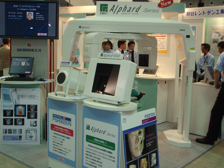
Figure 3
|
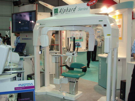
Figure 4
|
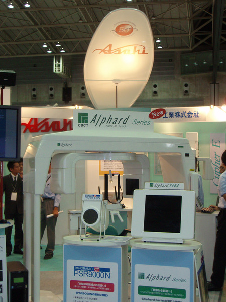
Figure 5
Dental CBCT systems can be classified into two categories as follows.
Dental (dento-alveolar): The FOV ranges approx. 5 cm (2 inch) to 10 cm (4 inch)in diameter. The isotropic voxel size ranges 0.1 mm to 0.2 mm.
Facial (maxillo-facial): The FOV ranges approx. 10 cm (4 inch) to 20 cm (8 inch) in diameter. The isotropic voxel size ranges 0.3 mm to 0.4 mm.
The "Alphard" having the largest FPD has the capacity to take CT images with any of these fields of view.
During the Autumn Meeting of the Japanese Society of Medical Imaging and Information Sciences in Tokyo, on October 7, 2006, I requested information on Alphard image quality from Prof. N. Nakamori, Kyoto Institute of Technology (http://www.mii-sci.jp/). He and his students are working with Asahi's CBCT images and at that meeting their presentations included "image noise removal using Wavelet transform and Sobel filter" and "recovery/improvement of image degradation at peripherals of FOV caused by localized image reconstruction." With objects having relatively higher/lower X-ray transparency are located on the contour of the localized FOV and partly included in the FOV, this causes specific artifacts; however, these artifacts can be minimized given optimal procedure settings.
New CBCT System (2): "FineCube"
Illustrations of "FineCube" manufactured by Yoshida Dental Manufacturing (Tokyo) are found in Fig. 6-Fig. 8 (http://www.yoshida-dental.co.jp/02/02_05.htm). The first exhibition of this system in Japan was carried out during the International Dental Show in Yokohama, Japan in October, 2006. "FineCube" was also exhibited by TeraRecon, Inc., at the ADA Annual Session in Las Vegas (http://www.terarecon.com/) and has previously in 2005 (and subsequently in 2006) was exhibited at two consecutive meetings of RSNA in Chicago. FDA approval for sale and use is pending in the USA. TeraRecon was established at Silicon Valley, CA, USA, by a Japanese entrepreneur and has a major office also in Chiba, Japan. Key products, which are both the 3D image-processing workstation and 3D-volume-rendering hardware, are well-known in Japan. There is the artist's rendering of the system at: http://www.auntminnie.com/index.asp?sec=road&sub=cto_2005&pag=dis&itemId=68307
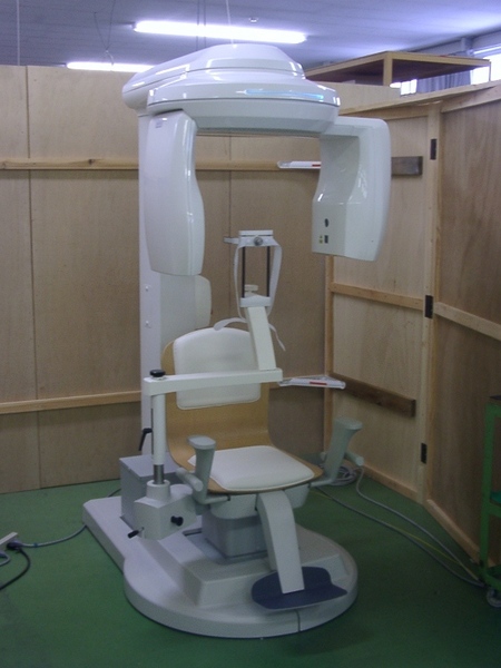
Figure 6
|

Figure 7
|
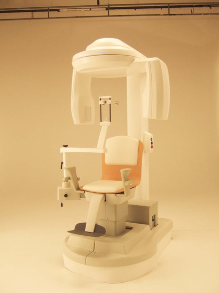
Figure 8
The chair and head-positioner look similar to those of Hitachi's CB MercuRay/CB Throne. The position in the dental market is an inexpensive and high-cost-performance CBCT unit for limited to medium size FOVs for implantologists compared with Hitachi's CBCT units. The two available FOV (diameter x height) settings are 56.5x51.7 mm and 82.0x75.1 mm. Two alternatives of exposure time setting for 360-degree rotation are 19 and 37 second. The choice will affect on image quality and exposure.
One good specification is the small footprint of the main body. The minimum requirement is only 1500x1800 mm (5 ft. x 5.6 ft.). Also the required lead shield is 1.5mmPb. All X-ray examinations in dental office are carried out in the proprietary room for exclusive X-ray use in Japan. Since the possession ratio of rotational panoramic radiography machines is more than 90%, two X-ray machines, namely dental and panoramic units, are installed in such a special room in most Japanese dental offices. Usually the lead protection of walls is 1.5mmPb in such cases. Hence, in Japan, the FineCube can be placed in a regular dental office without major reconstruction.
Additionally, the effective focal-spot size, 0.2 mm x 0.2 mm is surprisingly small. The business of "FineCube" is just under way; therefore details of performance are not yet available.
New CBCT System (3): "CB Throne"
The CB Throne, the 2nd CBCT system of Hitachi Medical Corp., was announced at April 2005. (Fig. 9) The sales started in fall 2005, but it's still focused only on the Japanese domestic market. The CB Throne is discussed at: http://www.hitachi-medical.co.jp/product/cbct/detail.html but the details are in Japanese.
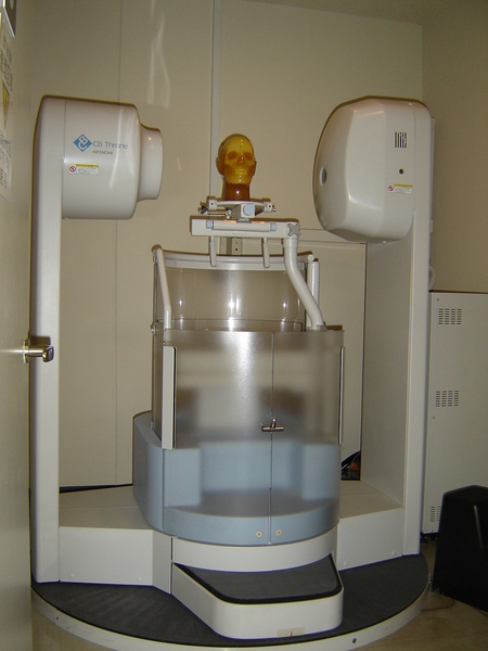
Figure 9
In comparison with CB MercuRay, since a 7-inch image-intensifier is installed, only the D-mode (sphere FOV: 5 cm in diameter, Slice: 0.1 mm thick) and I-mode (FOV: 10 cm in diameter, Slice: 0.2 mm thick) are available. Both P-mode and F-mode of CB MercuRay are not available. A clinical study has been made using the CB Throne at Tokyo Dental College Chiba Hospital. Our report written in Japanese can be downloaded at MEDIX site; http://www.hitachi-medical.co.jp/medix/. [file "P23-26.pdf (411kb)," at Vol. 44 articles.]
The power of the metal artifact reduction function is shown in Fig. 10. This was taken by the I-mode using the CB MercuRay and processed by 3D-reconstruction and rendering software. Usually severe metal artifacts are observed in the case when the medical CT examination and can be reduced significantly with the Hitachi CBCT algorithms.
The power of the metal artifact reduction function is shown in Fig. 10. This was taken by the I-mode using the CB MercuRay and processed by 3D-reconstruction and rendering software. Usually severe metal artifacts are observed in the case when the medical CT examination and can be reduced significantly with the Hitachi CBCT algorithms.
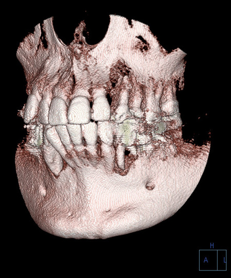
Figure 10
New Computed Radiography System: "YCR-21XG Bellissimo"
The "YCR-21XG Bellissimo" manufactured by Yoshida Dental Manufacturing (Tokyo) and sold by CrossField (Tokyo) is a new photostimulable phosphor computed radiography system for the dental office. Illustrations of this system were made at the private dental office of Dr. Y. Otake, Chiba, Japan. (http://www17.plala.or.jp/cosmosdental/ Fig. 11-Fig. 13) Dr. Y. Otake, with whom I have a lot of collaborative works, was purchased it in 2004. Her satisfaction with the image quality and operational performance are good.
The "YCR-21XG Bellissimo" manufactured by Yoshida Dental Manufacturing (Tokyo) and sold by CrossField (Tokyo) is a new photostimulable phosphor computed radiography system for the dental office. Illustrations of this system were made at the private dental office of Dr. Y. Otake, Chiba, Japan. (http://www17.plala.or.jp/cosmosdental/ Fig. 11-Fig. 13) Dr. Y. Otake, with whom I have a lot of collaborative works, was purchased it in 2004. Her satisfaction with the image quality and operational performance are good.
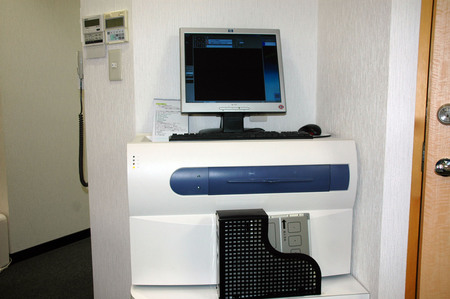
Figure 11
|
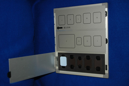
Figure 12
|
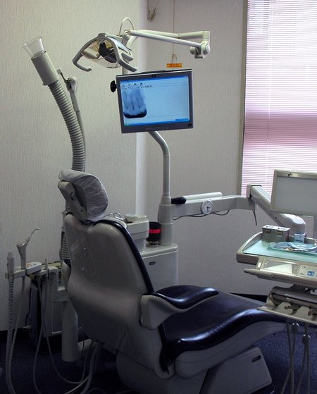
Figure 13
Important concepts and specifications of YCR-21XG are:
(1) To avoid the visible light effect to PSP (photostimulable storage phosphor) imaging plates YCR-21XG enables opening of cassette for panoramic and cephalometric radiography examinations automatically, ensuring that there is no possibility that PSP plates are exposed to visible light. In the case of intraoral radiography, also exposure to visible light exposure is limited.
(2) Logarithmic conversion from analogue data to digital signal: To maximize the X-ray contrast at any depth, YCR-21XG takes the logarithmic conversion of analog data, namely photo-intensities at each pixel measured by laser-stimulation, to digital data, namely pixel values which directly affect on the gray level display. Such logarithmic conversion is taken on most of medical CR scanner units. The benefit is to keep high contrast at both relatively low and high gray level regions on each image. The drawback is the increase of image noise.
To my knowledge, technical assessments and clinical studies have never been published on academic journals written in English. Nevertheless, the reader is referred to an article written by Dr. Predrag Sukovic in the Library, www.aadmrt.com"The monochromatic X-rays in homogenous objects show the logarithmic attenuation he described."
Summary
Taking this opportunity, I introduced three cone-beam CT units and a CR unit (and JORT). They all are available in Japan, but not elsewhere. I have no idea of the future availability of these units in detail in the United States and other countries on the globe.
REFERENCES
- Arnheiter C, Scarfe WC, Farman AG. Trends in maxillofacial cone-beam computed tomography usage. Oral Radiology (Japan) 2006; 22: 80-85.
- My submissions to the ORADLIST mailing lists at 03 Oct 2006, 08 Sep 2006, 16 Nov 2005, etc.
- Sukovic P. Cone Beam Computed Tomography in Dentomaxillofacial Imaging. in the Library, www.aadmrt.org