
NewTom 9000David C. Hatcher, DDS, MSc, MRCD (c)

Images of the craniofacial region comprise an important component of the dental patient record. Ideally, the imaging process begins with the development of an imaging goal. The imaging goal is a clinically derived question that employs imaging to find the solution, in other words, using imaging as a tool to seek the answer to a clinical question. Specific and detailed clinical questions require detailed and specific imaging solutions. Digital processes have improved the diagnostic capabilities of the imaging tools being used in dentistry.
DIGITAL IMAGE CHARACTERISTICS: A digital image is composed of picture elements (pixels) that are arranged in a 2-dimensional rectangular grid with each pixel having a specific size, color and intensity value and location within the image (ie., bitmapped [BMP] or raster). A pixel is the smallest element of a digitized image. Radiographic images generally use a gray color with an intensity value between 8 bits (28 or 256 shades of gray) and 12 bits (212 or 4096 shades of gray). Image resolution is the degree of sharpness of an image. The image resolution is influenced by the number of pixels per given length of an image, (i.e. pixels/mm), the number of gray levels per pixel (bits) and the management of the gray levels. Selected digital imaging devices can produce digital volumes or 3-dimensional images. The volume element (voxel) is the smallest element of a 3-dimensional image. A voxel volume can be thought of as a 3-dimensional array or stack of bitmapped images with each voxel having a height, width and thickness. 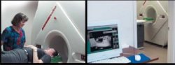
FIGURE 1: The images show the NewTom 9000 Volume Scanner from Verona, Italy. The left image shows Camille Mayorga, DRT, talking to a patient prior to entry into the scanner while the right image shows the adjacent computer workstation. Courtesy of Diagnostic Digital Imaging, Sacramento, CA.
|


|
NEW DIGITAL IMAGING DEVICES: New and future imaging trends for dentistry include digital imaging and 3-dimensional imaging of the maxillofacial regions. The ultimate reward of the technological imaging advancements is the digital representation of the patient's anatomy as it exists in nature (anatomic truth). Multiplanar reformatting of the accurate digital 3-dimensional image data volume using software tools can provide clinically relevant diagnostic and spatial information. A digital imaging breakthrough, the NewTom 9000 volume scanner, is now available for clinical practice.
NewTom 9000: The volume imaging technique employs the principle of tomosynthesis and is also known as cone beamed CT. The NewTom 9000 (Mozzo, et. al.), a volume imaging machine produced in Italy, received FDA approval in April 2001 (Figure 1). The NewTom 9000 has been designed specifically to image the maxillofacial region. In a single scan the x-ray source and a reciprocating x-ray sensor rotate around the head and acquire 360 pictures (one image per degree of rotation) using 17 seconds of accumulated exposure time. The entire maxillofacial volume (13 cm diameter field of view) is imaged and the patient receives an absorbed dose similar to a periapical survey of the dentition. The 360 acquired images undergo a primary reconstruction to mathematically replicate the patient's anatomy into a single 3D volume that is comprised of volume elements (voxels) similar to a Rubik's cube. Each voxel is small (0.29 mm for each of the cube faces) and therefore the image has a relatively high resolution. The NewTom software allows for reformatting and viewing the image data from any point of view in straight or curved planes and in 3-D (Figures 2,3,4). Using these software tools the anatomy can be peeled away layer by layer to locate the desired anatomy. The NewTom 9000 scanner ranks extremely high when considering the balance between high diagnostic yield, low cost and low risk.
NewTom 9000: The volume imaging technique employs the principle of tomosynthesis and is also known as cone beamed CT. The NewTom 9000 (Mozzo, et. al.), a volume imaging machine produced in Italy, received FDA approval in April 2001 (Figure 1). The NewTom 9000 has been designed specifically to image the maxillofacial region. In a single scan the x-ray source and a reciprocating x-ray sensor rotate around the head and acquire 360 pictures (one image per degree of rotation) using 17 seconds of accumulated exposure time. The entire maxillofacial volume (13 cm diameter field of view) is imaged and the patient receives an absorbed dose similar to a periapical survey of the dentition. The 360 acquired images undergo a primary reconstruction to mathematically replicate the patient's anatomy into a single 3D volume that is comprised of volume elements (voxels) similar to a Rubik's cube. Each voxel is small (0.29 mm for each of the cube faces) and therefore the image has a relatively high resolution. The NewTom software allows for reformatting and viewing the image data from any point of view in straight or curved planes and in 3-D (Figures 2,3,4). Using these software tools the anatomy can be peeled away layer by layer to locate the desired anatomy. The NewTom 9000 scanner ranks extremely high when considering the balance between high diagnostic yield, low cost and low risk.
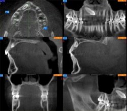
FIGURE 2: This image collage shows multiplanar reformation of the NewTom 9000 Volume data to visualize the facial anatomy and impacted tooth #6. The top left image displays the maxillary anatomy in the axial plane. The top right image displays the anatomy in a curved plane similar to a panoramic projection. The middle two images show sagittal sections of the head near the midline. In this section the coupling of the anterior teeth, hard and soft palate, tongue and pharyngeal air space can be visualized. The bottom left image shows an axial section through the molar teeth, maxillary sinuses, nasal fossa and mandible. The bottom right image is an axially corrected view of the right TMJ while visualizing the teeth in occlusion. Courtesy of Diagnostic Digital Imaging, Sacramento, CA.
The patient image data can be organized into a "mounting template" and viewed on the computer screen, copied to a floppy disk or CD ROM or printed using a diagnostic quality print in glossy paper or transparency formats. This is similar to the way that digital extra and intra oral photographs are mounted. In addition, the entire data volume can be exported to a CD ROM in DICOM format and transferred to any computer to be reconstructed and viewed as shown in Figures 2-4 using software tools that are available for distribution to the dental community.
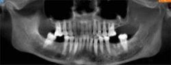
FIGURE 3: This image is reformatted in a curved plane that has a buccolingual thickness of 12 mm. Courtesy of Diagnostic Digital Imaging, Sacramento, CA.
CONCLUSIONS: Computer assisted imaging is now creating the opportunity for the dental profession to better visualize and study the craniofacial anatomy. New imaging tools like the NewTom 9000 allow for accurate 3-dimensional replication and display of the patient in the form of voxel volumes. There are interactive software tools that allow the clinician to peel away the tissue layers and reveal the hidden anatomy.NewTom 9000 Volume scan has been extremely valuable for the investigation of impacted teeth, temporomandibular joints, implant planning and pathology. At this time the NewTom 9000 volume scans occasionally need to be supplemented with panoramic, cephalometric and periapical projections.
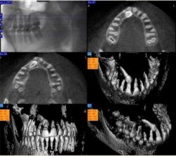
FIGURE 4: This image collage has been designed to visualize impacted tooth #6 in the axial plane (upper right and middle left sections) and in 3 dimensions. This type of visualization can be utilized to spatially determine the location of the impacted tooth relative to the roots of the adjacent teeth. Courtesy of Diagnostic Digital Imaging, Sacramento, CA.
REFERENCES
Mozzo P, Procacci C, Tacconi A, Tinazzi Martini P, Bergamo Adnreis, IA, A new volumetric CT machine for dental imaging based on the cone-beam technique: preliminary results. Eur Radiol 1998; 8:1558-1564