
Effective Dose of Two Cone-Beam CT Scanners:
|


|

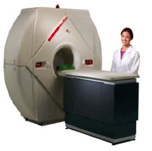
Figure 1
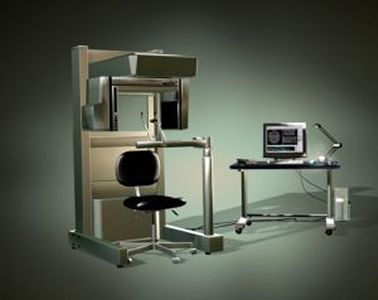
Figure 2
|
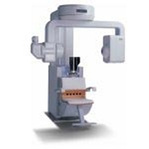
Figure 3
|
The dosimetry study was done on all scanners by inserting 24 TLDs (Landauer, Inc., Glenwood, IL) into the same RANDO phantom (Nuclear Associates, Hicksville, NY) and making 3 replications of a full field of view (FOV) scan to assure adequate exposure of the dosimeters. Dosimeter locations (number of TLDs) included the calvarium (3), mid brain (1), pituitary (1), orbits (2), lens of eye (2), cheek (1), parotid (2), ramus (2), cervical spine (1), back of neck (1), mandible body (2), submandibular glands (2), sublingual glands (1), thyroid (2), and esophagus (1). Average tissue-absorbed dose, weighted equivalent dose, and effective dose were calculated for each major anatomical site. Effective doses of individual organs were summed using tissue-weighting factors for both 1990 and proposed 2005 ICRP3 guidelines to calculate two measures of whole-body effective dose.
The major differences in the tissue-weighting factors between the 1990 and the proposed 2005 ICRP guidelines include the handling of the dose to the salivary glands, the brain and the remainder organs. The 2005 guidelines have a specific weight for salivary glands and the brain, whereas the 1990 guidelines did not. Since the salivary glands are in the primary beam during head CT, including their dose increases the validity of the calculation of effective dose with respect to actual patient risk.
For the full FOV I-CAT scan (see figure 4), the effective dose was 68.7 µSv using the 1990 tissue weights and 101.5 µSv using the 2005 tissue weights. The operating parameters were 120 kVp, 22.85 mAs. The effective doses for the NewTom 3G, using the same phantom and full FOV (see figure 5) were 43.1 and 56.5 µSv, for the two weighting factors, respectively. The operating factors were 110 kVp, 8.1 mAs as determined automatically after a prescan of the phantom.
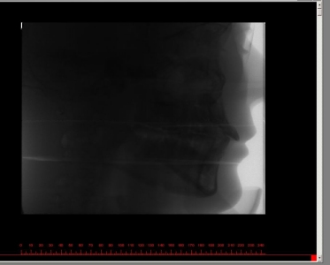
Figure 4
|
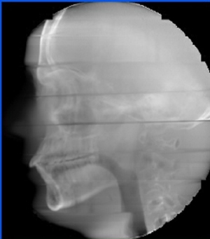
Figure 5
|
To put the doses from the CBCT scanners into perspective, published effective doses from digital panoramic radiography range from 4.7 to 14.9 µSv per scan.4 Other published data on non-digital panoramic radiographs puts the effective dose as high as 26 µSv. 5 Depending on which machine it is compared with, one full FOV I-CAT scan is the equivalent of 4-22 panoramic radiographs. The same text cited for the panoramic dose5 lists the effective dose for a 19-film complete mouth intraoral survey made with round collimation and D-speed film as 150 µSv, more than a single I-CAT (or NewTom) scan.
|
While the effective dose from the I-CAT is 1.6- 1.8 times that of the NewTom 3G, depending on which weighting factors are used, there is another part of the story that has not yet been determined: the issue of image quality vs. radiation dose. The operating parameters for the I-CAT are generally not adjustable by the user, although there are some software modifications that could be made that would allow lower kVp and mAs to be used, while those of the NewTom 3G are adjusted automatically based on a prescan. For the RANDO phantom imaged there was a difference of 10 kVp and 14.8 mAs between the two scans. Before the dosimetry data are published in a peer-reviewed journal, an attempt will be made to correlate the radiation dose to the image quality.
|
|
If the quality of the lower dose NewTom 3G images is as good as that of the higher dose I-CAT, perhaps adjustments could be made to the I-CAT to reduce the dose further beyond what has already been done with the fewer basis images and shaped beam. There may also be situations where image quality (and dose) could vary with the diagnostic task.
As the developers and manufacturers of cone-beam CT scanners continue to improve both the hardware and software of their equipment, there will be many changes in both image quality and radiation dose that will be of benefit to the users of the equipment and their patients.
As the developers and manufacturers of cone-beam CT scanners continue to improve both the hardware and software of their equipment, there will be many changes in both image quality and radiation dose that will be of benefit to the users of the equipment and their patients.
REFERENCES
- Ludlow JB, Davies-Ludlow LE, Brooks SL. Dosimetry of two extraoral direct digital imaging devices: NewTom cone beam CT and Orthophos Plus DS panoramic unit. Dentomaxillofacial Radiology 2003; 32: 229- 234.
- ICRP Publication 60. Radiation protection. 1990 recommendations of the International Commission on Radiological Protection. Ann ICRP 1991; 21.
- 2005 recommendations of the International Commission on Radiological Protection. Draft for consultation.http://www.icrp.org/docs/2005_recs_CONSULTATION_Draft.pdf
- Gijbels F, Jacobs R, Debaveye D, Verlinden S, Bogaerts R, Sanderink G. Dosimetry of digital panoramic imaging. Part 1: patient exposure. Dentomaxillofac Radiol 2005; 34: in press
- Frederiksen NL. Health physics. In, White SC, Pharoah MJ. Oral radiology: principles and interpretation, 5th ed. St. Louis: Mosby, 2004. page 54.