
The New Digital Age of Clinical PhotographyDr. Robert Shorey
Enhanced Dental Concepts Roseville, California From the Fall 2007 AADMRT Newsletter  Dr. Robert Shorey Dr. Robert Shorey
Introduction:
From its beginnings in the 1840's Clinical Photography has been recognized as an important medical documentation modality. Until recently, the expense and time of film development limited the widespread adoption of photography as a regular fundamental diagnostic tool in most everyday general dentistry offices. Digital photography has eliminated the lag time of film development and brought the capture of clinical images into the moment and minimized the guesswork in achieving a quality final image product. Clinical single lens reflex (SLR) cameras capable of producing distortion free, well focused images are affordable and substantially less expensive than previously favored wand-type intra-oral video cameras. In 2007 a quality SLR clinical photography camera can be assembled for under $2000. With training the versatility of these cameras allow for high resolution full facial to close-up 1:1 images of the teeth and oral structures chronicling care before, during and after dental treatment. Digital clinical photographic images are becoming an important mainstream diagnostic and communication tool to share with patients, lab technicians and colleagues. The ability to produce high resolution images at the time of the patient's clinical examination visit allows dentists to share their findings with patients as never before. When a dentist is reviewing a patient's comprehensive needs and cosmetic desires, digital photography is helping to narrow the gap between subjective patient expectations and clinical realities. Patients literally become part of the diagnostic process; seeing with their own eyes the interrelationship of factors that combine to make up their oral health. If cosmetic procedures are desired, patients can often come to a better understanding of the true clinical necessities that will allow them to achieve appropriate lasting cosmetic improvements. When comprehensive treatment plans are formulated, the information is easily shared through images with lab technicians and specialists assisting in the patient's care. |


|
Digital Clinical Photography Equipment:
The right equipment, training and the adoption of a regimented structured series of clinical images is necessary to gain full advantage of this technology. There are established resources like the Institute of Medical Illustrators or the American Academy of Cosmetic Dentistry that provide written guidelines for a structured series of clinical images. No matter the resource, these photographic series usually include several portrait images and several intra-oral images comprising anterior, occlusal and lateral views of the patient's dentition. Through these images the patient's skeletal form, symmetry, arch form, occlusion and arch relationships, tooth rotations and trajectories can be analyzed. A more robust appreciation for the patient's oral condition is obtained when this information is added to their radiographic and plaster study model information.
Digital cameras are constantly being upgraded through extensive competition for market dominance. An SLR clinical camera system consists of three components: Camera body, lens and macro flash system. As of the writing of this article (2007) recommendations for a quality clinical digital camera system would be the following:
Nikon D40/D40X (camera body), 6 MegaPixel/10MegaPixel SLR with Sigma 105mm Macro (lens), Sigma EM-140DG (ring flash)
Alternative options for lens and flash for Nikon D40 series are Nikkor 105mm Micro and Nikon R1C1 flash system.
Canon XTi (camera body), 10MegaPixel SLR with Sigma 105mm Macro (lens), Sigma EM-140DG (ring flash)
Alternative Camera Body is the Canon 40D (camera body) 10MegaPixel SLR
Alternative options for lens and flash for Canon XTi are Canon 100mm Macro lens and Canon MR14EX ring flash system.
These systems would be considered excellent entry level quality image systems. More sophisticated camera body systems would be the Canon 5D or Nikon D200 or soon to be released Nikon D300.
Note: When choosing a camera system, resolutions of 6MegaPixel or greater will produce high quality clinical images. A clinical camera is composed of camera body, lens and ring flash. The camera lens is the most likely determining factor of what brand of camera body you will choose. Future camera upgrades are often dependent on lenses which are proprietary to specific camera body brands.
Application of Structured Clinical Image Information:
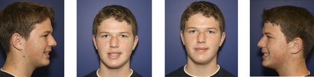
Figure 1. Typical clinical facial photography series
Through the use of a structured photographic routine similar to a clinical radiographic series consistent image information can be gathered that will provide important clinical diagnostic findings. The portrait series of images usually consists of at least 3-4 views (frontal smile and frontal lip at rest, right and left lateral). These image views provide important skeletal, facial symmetry and smile display information. This type of information can not be adequately achieved through any other diagnostic modality such as study models.
Captured portrait images allow for quantitative analysis of the smile and facial symmetry. Using programs like Adobe Photoshop an image can be measured for symmetry using lines and equivalent size boxes such as the one in figure 2. The subject in figure 2 does not require any dental services and has an appealing healthy smile. Notice the asymmetries illustrated when studying her photographic image. The lines and boxes allow us to more clearly evaluate the relative spacing of anatomic features. In figure 2 the eyes, nose and dental midline can not be completely centered. Following along, the subject in figure 3 does require treatment. If the challenges of asymmetries are not revealed before treatment begins this person, husband or their parents might be unhappy with the final achievable results. Study models without clinical photos would not provide sufficient evidence that the midline of her nose and upper arch are not unified. Sharing these images with the patient and co-treatment specialists can provide a clear road map to necessary adjunctive treatment needs such as orthodontic tooth movement.
Captured portrait images allow for quantitative analysis of the smile and facial symmetry. Using programs like Adobe Photoshop an image can be measured for symmetry using lines and equivalent size boxes such as the one in figure 2. The subject in figure 2 does not require any dental services and has an appealing healthy smile. Notice the asymmetries illustrated when studying her photographic image. The lines and boxes allow us to more clearly evaluate the relative spacing of anatomic features. In figure 2 the eyes, nose and dental midline can not be completely centered. Following along, the subject in figure 3 does require treatment. If the challenges of asymmetries are not revealed before treatment begins this person, husband or their parents might be unhappy with the final achievable results. Study models without clinical photos would not provide sufficient evidence that the midline of her nose and upper arch are not unified. Sharing these images with the patient and co-treatment specialists can provide a clear road map to necessary adjunctive treatment needs such as orthodontic tooth movement.
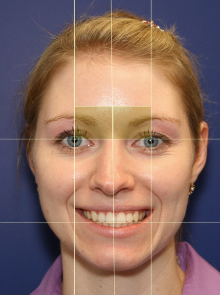
Figure 2. Full facial portrait image
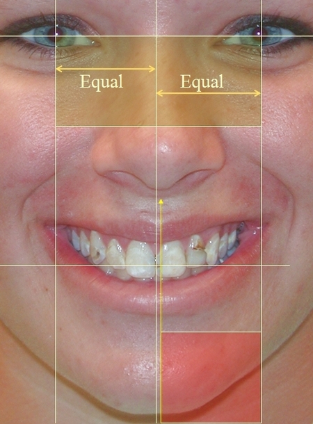
Figure 3. Facial view with asymmetry noted from nose midline
Intra-oral clinical views often consist of anterior, occlusal and lateral views of the dentition. These images can provide a multitude of diagnostic information ranging from visible caries, cracks, occlusal/incisal wear, tooth positions, arch relations and arch form. A well imaged occlusal view is a valuable communication resource and provides clear details and easily recognizable relationships of the teeth. As illustrated in figure 4, the broken tooth that brought the patient in to seek treatment is just the tip of the iceberg concerning their dental health problems that need professional remedy.
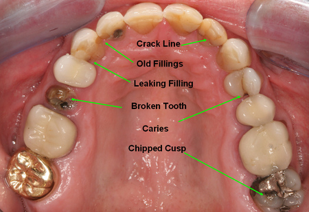
Figure 4. Maxillary occlusal view
In figure 5 the patient sought treatment for his lower front tooth. Using an occlusal view of the lower arch a discussion with the patient about the appearance of his back teeth resulted in seeking care and diagnosis from his physician of gastro-esophageal reflux disease (GERD). The image provided evidence of acid attack of enamel surfaces.
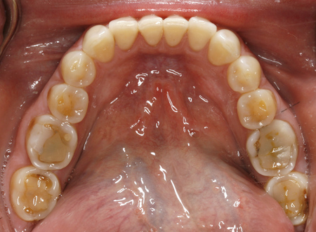
Figure 5. Mandibular occlusal view
Figure 6 illustrates a serious crack that can compromise the long term prognosis of the molar tooth and may necessitate root canal therapy. Figure 7 also illustrates a reason for possible root canal treatment if the tooth is crowned to raise the occlusal table to harmonize with the adjacent plane of occlusion. Review of a photographic image compared to a radiograph can illustrate the closeness to the pulp chamber after tooth reduction for the crown.
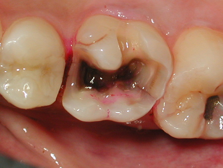
Figure 6. Cracked Molar
|

Figure 7. Extruded Molar
|
Such visual results can predict the likely need for endodontic treatment before treatment. Providing this information to a referral doctor would minimize any possible misunderstandings when they confer with the patient about treatment objectives and prognosis.
Dental conditions like periodontal disease and poor oral hygiene can be elucidated through captured visuals of the patient's plaque control. Notice the plaque build-up on the teeth in figure 8. A photograph of the disclosed teeth in figure 9 is an even more striking illustration of the amount of plaque. Pictures taken at a later time can be used to illustrate progress in the patient's oral hygiene visits and will not only show less plaque but can show improved gum health through evidence of firmer tissue and less inflammation.
Dental conditions like periodontal disease and poor oral hygiene can be elucidated through captured visuals of the patient's plaque control. Notice the plaque build-up on the teeth in figure 8. A photograph of the disclosed teeth in figure 9 is an even more striking illustration of the amount of plaque. Pictures taken at a later time can be used to illustrate progress in the patient's oral hygiene visits and will not only show less plaque but can show improved gum health through evidence of firmer tissue and less inflammation.
Figure 8. Teeth with heavy plaque
Figure 9. Disclosed teeth with plaque
Another example of disease recorded through imagery is in figure 10 and 11. The irrefutable communication power of images convinced this patient to stop wearing a metal stud. The evidence of the damage the stud was doing to the gum tissue was easily shown to this young 20 year old patient.
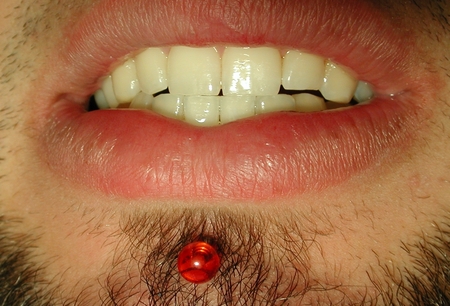
Figure 10. Jewelry lip stud with metal back
|
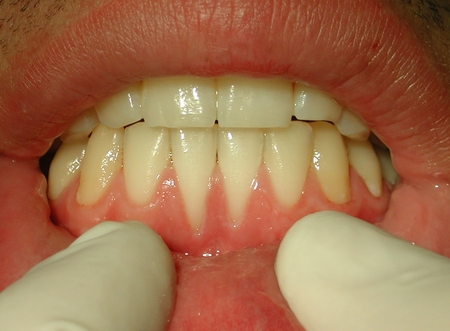
Figure 11. Recession from stud's metal back
|
Digital photography is not just for communication with patients and specialists. Clinical images allow lab technicians assisting in a patient's care to have a better understanding of treatment objectives and challenges.
In figure 12 a neutral gray card is utilized to block out distractions and color correct the image for improved image color fidelity. Surface texture and color gradations can be communicated to the dental laboratory using digital photography. When ceramic restorations are being fabricated this additional information helps to improve the ceramic technicians ability to produce cosmetic harmony of crowns with the adjacent natural dentition.
In figure 12 a neutral gray card is utilized to block out distractions and color correct the image for improved image color fidelity. Surface texture and color gradations can be communicated to the dental laboratory using digital photography. When ceramic restorations are being fabricated this additional information helps to improve the ceramic technicians ability to produce cosmetic harmony of crowns with the adjacent natural dentition.
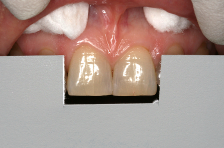
Figure 12. Anterior teeth with gray photo gray card
Technicians seldom meet patients for whom they provide laboratory services. Images help them understand the human aspect of their work and give them an appreciation for the final results. In figure 13 the challenge of creating a life-like result for a dental implant case can be better appreciated through clinical images and the ultimate results more often reach their mark when the lab technician's artistry is applied to meet the visual and biologic challenges.
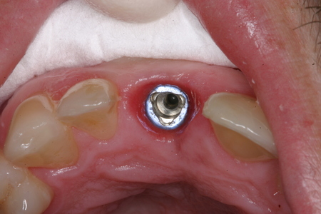
Figure 13. Anterior implant
|
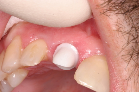
Figure 14. Secondary abutment
|
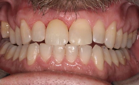
Figure 15. Same day crown seated
|
Conclusion:
The production of clinical images through digital SLR photography is more affordable and faster than ever. The ability to gain almost instant image results allows clinical photographic images to be readily perfected and shared with patients, lab technicians and co-therapists. Regimented clinical photography adds substantial diagnostic information to the established regimens of study models and dental radiographs. A picture is worth a thousand words providing an efficient and persuasive communication and documentation tool. The affordability and utility of digital photography is quickly making it an expected part of the standard of dental care.
The production of clinical images through digital SLR photography is more affordable and faster than ever. The ability to gain almost instant image results allows clinical photographic images to be readily perfected and shared with patients, lab technicians and co-therapists. Regimented clinical photography adds substantial diagnostic information to the established regimens of study models and dental radiographs. A picture is worth a thousand words providing an efficient and persuasive communication and documentation tool. The affordability and utility of digital photography is quickly making it an expected part of the standard of dental care.