
Exploring the Mandibular Canal in 3 Dimensions: An Overview of Frequently Encountered Variations in Canal AnatomyDr. Shaza Mardini and Dr. Anita Gohel
From the Fall 2008 AADMRT Newsletter
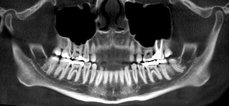
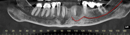
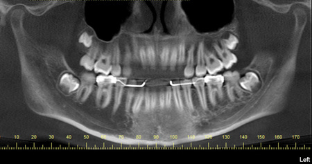
Figure 1: A reconstructed panoramic image displayed in a thin section to show the bilateral course of the mandibular canals. The canal on the right has been indicated by the white arrows
|


|


Variation of normal anatomy:
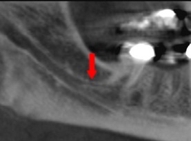
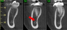
Common variations are seen with respect to the anatomy and location of the mandibular canal. One such anatomic variation is known as a bifid mandibular canal. The canal may be split higher up near the foramen or other variations may be seen where the canal splits off in different positions. Often one canal may be smaller than the other. It has been reported in the literature that there is 0.95% prevalence of bifid mandibular canals (Langlais et al). Figure 2 shows some examples of a bifid canal.
Common variations are seen with respect to the anatomy and location of the mandibular canal. One such anatomic variation is known as a bifid mandibular canal. The canal may be split higher up near the foramen or other variations may be seen where the canal splits off in different positions. Often one canal may be smaller than the other. It has been reported in the literature that there is 0.95% prevalence of bifid mandibular canals (Langlais et al). Figure 2 shows some examples of a bifid canal.
2a
2c
|

2b
2d
|
Figure 2: (a) shows a panoramic view of a bifid canal (b) in cross section one smaller canal (red arrow) superior to a larger canal(white arrow) (c) shows the canal splitting off higher up near the ramus (d) the smaller canal is seen superior in cross section (red arrow) and the main canal inferior (white arrow).
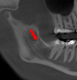
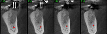
In addition to the bifid canal, a double mental foramen may be seen (figure3). Another anatomic variation of the canal which we encounter frequently when viewing the mandible in three dimensions is the anterior loop or incisive branch of the mandibular canal. This represents a branching of the mandibular nerve anterior to the mental foramen with a continuation or extension of the canal seen. It has been reported in the literature with prevalence in 7% of cases (Jacobs et. Al, 2002). This anatomical variation can complicate mandibular anterior implant placement because the canal houses neural and vascular tissue which should be avoided. The anterior loop is often well visualized in cross sectional views great attention should be paid to this variation. Figure 4 shows an example of an anterior loop of the mandibular canal/mental foramen.
In addition to the bifid canal, a double mental foramen may be seen (figure3). Another anatomic variation of the canal which we encounter frequently when viewing the mandible in three dimensions is the anterior loop or incisive branch of the mandibular canal. This represents a branching of the mandibular nerve anterior to the mental foramen with a continuation or extension of the canal seen. It has been reported in the literature with prevalence in 7% of cases (Jacobs et. Al, 2002). This anatomical variation can complicate mandibular anterior implant placement because the canal houses neural and vascular tissue which should be avoided. The anterior loop is often well visualized in cross sectional views great attention should be paid to this variation. Figure 4 shows an example of an anterior loop of the mandibular canal/mental foramen.

Figure 3: 3D volume rendering of a mandible with two mental foramina.
4a
4b
Figure 4:(a) Shows a reconstructed panoramic at 5mm thickness, the mandibular canal on the left has been traced in red. Note the anterior extension of the mental foramen (b) Images of the same patient shown in cross section cross referenced to the panoramic. Note the mental foramen at slice #21 and the continuation of the canal anteriorly to the midline.
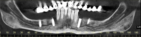
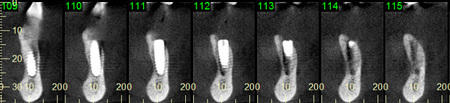
Figure 5:(a) A reconstructed panoramic view with implants placed in the partially edentulous mandible. The implant seen distal to #22 has penetrated the anterior loop of the mandibular canal. Note the implants on the right are both in the canal as well. (b) Cross sectional images of the implant distal to tooth #22.
Pathology of the Mandibular Canal
The mandibular canal houses neural and vascular tissue and therefore is subject to pathology associated with these tissues. Neural tumors such as neuromas, neurofibromas, schwannomas and neural malignancies are seen in the mandibular canal. In addition, vascular tumors are also seen associated with the canal such as A-V malformations, hemangiomas and malignancies such as lymphomas.
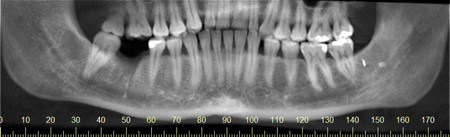
These types of pathology are often detected by the change in size, shape or location or course of the mandibular canal. For example, with the neural tumors there is often an abnormal widening of the mandibular canal seen in the location of the tumor (figure 6).
Pathology of the Mandibular Canal
The mandibular canal houses neural and vascular tissue and therefore is subject to pathology associated with these tissues. Neural tumors such as neuromas, neurofibromas, schwannomas and neural malignancies are seen in the mandibular canal. In addition, vascular tumors are also seen associated with the canal such as A-V malformations, hemangiomas and malignancies such as lymphomas.
These types of pathology are often detected by the change in size, shape or location or course of the mandibular canal. For example, with the neural tumors there is often an abnormal widening of the mandibular canal seen in the location of the tumor (figure 6).
6a
6b
Figure 6:(a): A reconstructed panoramic view of the mandible showing the right mandibular canal more narrow that the left. (b) In cross section the canal on the patient's left appears larger in all dimensions. The canal is seen next to the red arrows.
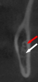
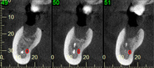
Vascular tumors may cause a change in position of the canal, superiorly or inferiorly and may cause it to appear tortuous (figure 7). Other pathology that can be seen are foreign bodies within and around the canal (figure 7).
Vascular tumors may cause a change in position of the canal, superiorly or inferiorly and may cause it to appear tortuous (figure 7). Other pathology that can be seen are foreign bodies within and around the canal (figure 7).
7a
7b
Figure 7:(a) Reconstructed panoramic (b) Note the foreign body was buccal to the canal. (c) On the left side the foreign body was seen at the superior border of the canal.
To summarize, the mandibular canal has a natural bilateral and symmetric course in the mandible. Variations in the anatomy are seen such as bifid canal, double mental foramen and anterior looping. Pathology of the canal is often detected by a change in size shape or course of the canal. Knowledge of these variations helps in diagnosis and treatment planning of procedures in the mandible and lead to fewer complications during treatment.
To summarize, the mandibular canal has a natural bilateral and symmetric course in the mandible. Variations in the anatomy are seen such as bifid canal, double mental foramen and anterior looping. Pathology of the canal is often detected by a change in size shape or course of the canal. Knowledge of these variations helps in diagnosis and treatment planning of procedures in the mandible and lead to fewer complications during treatment.
REFERENCES
Jacobs, R. Mraiwa, N. vanSteenberghe, D. Gijbels, F and Quirynen, M. Appearance, location, course, and morphology of the mandibular incisive canal: an assessment on spiral CT scan. Dentomaxillofacial Radiology (2002) 31, 322-327.
Langlais, R.P. Broadus, R. Glass, B. Bifid mandibular canals in panoramic radiographs. Journal of the American Dental Association (1985), Vol 110, Issue 6, 923-926.
Langlais, R.P. Broadus, R. Glass, B. Bifid mandibular canals in panoramic radiographs. Journal of the American Dental Association (1985), Vol 110, Issue 6, 923-926.