
Use of Panoramic Radiography in Private Orthodontic PracticeLee Mettler, D.D.S.
Sacramento State University, Bachelor's Deg. at U.C. Davis, UCLA School of Dentistry, Orthodontic Training at UCSF, He has been in practice for the last 33 years. From the summer 2010 AADMRT Newsletter  Dr. Lee Mettler Dr. Lee Mettler
Orthodontists have relied on panoramic radiography as a principle tool for orthodontic diagnosis and treatment planning since the use of panoramic radiographs became widespread during the 1970's and 1980's. Since that time, orthodontists have found additional applications of this highly useful and valuable tool for improved orthodontic treatment results.
FIGURE 1 below shows various orthodontic characteristics of an 11Y, 11M old female patient with 1) three missing teeth 2) a supernumerary tooth distal to the upper right lateral incisor 3) the upper left cuspid's forward eruption into the missing upper left lateral incisor position and 4) the supernumerary tooth with a conical shaped crown. Surprisingly, the four third molars appear normal for their stage of development and their positions for the age of the patient. 
Figure 1: INITIAL RECORDS 2-04-2009 Pano - (Patient #1)
|


|
Patient #1's clinical orthodontic condition is shown in the following photo. The upper right lateral incisor and the supernumerary crown are shown at early eruption.
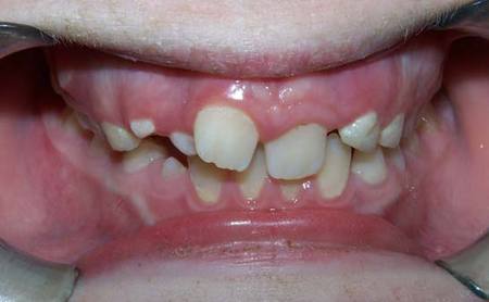
Figure 2: INITIAL RECORDS 5-21-2008 - (Patient #1)
FIGURE 3 below shows two years of eruption progress for Patient #1 This panoramic view shows eruption progress suitable for full braces placement.
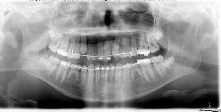
Figure 3: PHASE I PROGRESS (2) 5-13-2010 Pano - (Patient #1)
FIGURES 4 and 5 (clinical photos) below show two views of Patient #1's dentition about two months before the Panoramic view shown previously in FIGURE 3.
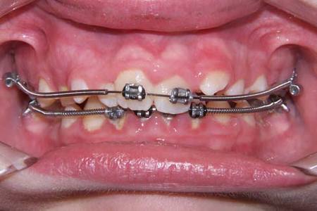
Figure 4: PHASE I PROGRESS (1) 3-10-2010 - (Patient #1)
|
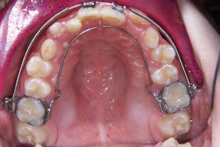
Figure 5: PHASE I PROGRESS (1) 3-10-2010 - (Patient #1)
|
Teeth vertical, parallel and evenly spaced is a goal desired during full and/or partial fixed appliances orthodontic treatment. Orthodontists expanded the application of panoramic view radiography by using panoramic's during finishing tooth movement. The following two cases show examples of initial and progress radiographs in the use of panoramic radiography for late stage finishing treatment.
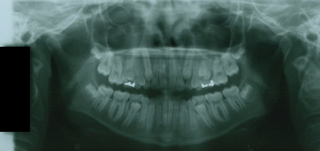
Figure 6: INITIAL RECORDS 1-09-2008 (Age 12-2) Pano - (Patient #2)
|
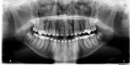
Figure 7: PROGRESS RECORDS 5-25-2010 (Age 15-1) (Patient #2)
|
The next case (Patient #3) shows both initial and late-in-treatment progress Pano's with the teeth in corrected and favorable positions for removal of teeth. Note the experienced root rounding of the apices of the upper and lower incisors shown in the 2nd panoramic.

Figure 8: INITIAL RECORDS 8-14-2007 (Age 11-11) Pano (Patient #3)
The lower left 2nd bicuspid (tooth #20) has its crown tipped to the distal and the root of the tooth in a mesially-tipped position. Adjusting the angle of the archwire at the portion of the wire for tooth #20 will improve the tooth to vertical and match the position of the lower right 2nd bicuspid. Note the patient experienced root rounding of the apices of the upper and lower central incisors seen in the May, 2010 Pano.
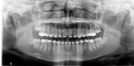
Figure 9: PROGRESS RECORDS 5-18-2010 (Age 14-8) (Patient #3)
Third Molar Relationship
Third molar teeth develop their roots during the traditional ages of full, fixed appliance treatment at ages 11 to 13 years old. Panoramic radiography is the most used form of examination for third molar diagnosis. Orthodontists in conjunction with general dentists and oral surgeons often find third molar conditions which indicate third molar removal before full, fixed appliance treatment begins.
FIGURES 10 and 11 shows an 11Y, 5M age patient in the early adult dentition. The upper and lower occlusal photos show three of the four 2nd molars through the gingival tissue. This is the classic appearance of a patient stage of dental development and eruption suited for beginning full, fixed appliance treatment.
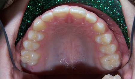
Figure 10: INITIAL RECORDS 3-12-2009 - Upper Occlusal Photo
|
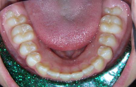
Figure 11: INITIAL RECORDS 3-12-2009 - Lower Occlusal Photo
|
The panoramic view shows an ectopic position of the UL 3rd molar at the occlusal surface of the UL 2nd molar. The normal eruption path of the UL 2nd molar is being blocked by the 3rd molar developing crown.
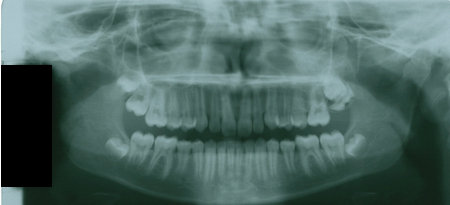
Figure 12: INITIAL RECORDS 3-19-2009 Panoramic View
The Lateral Cephalometric view shows the UL 2nd molar held up in position and delayed in eruption compared with the more normal UR 2nd molar:
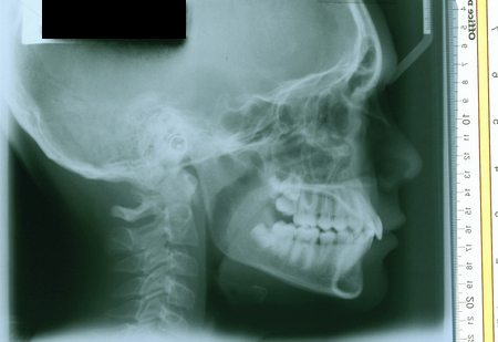
Figure 13: INITIAL RECORDS 3-19-2010 Lateral Cephalometric
The two periapical views below from the general dentist taken 3-27-09 and 6-18-09 show the eruption of the UL 2nd molar into the UL 3rd molar crown.
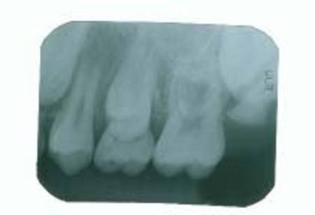
Figure 14: 3-27-2009 Periapical View
|
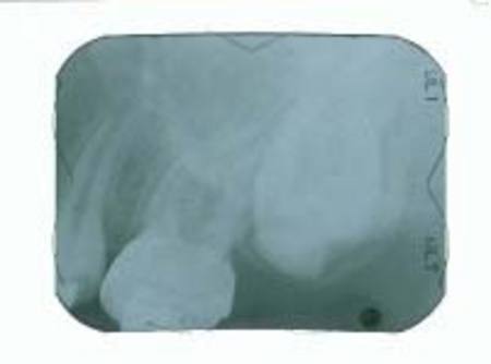
Figure 15: 6-18-2009 Periapical View
|
The ectopic UL 3rd molar was removed in July, 2009. The recent panoramic view below dated 4-5-2010 shows the 2nd molar able to proceed with unobstructed eruption but still about the same distance above the level of the contralateral UR 2nd molar (seen on the Lateral Cephalometric dated 3-19-2009 shown earlier).
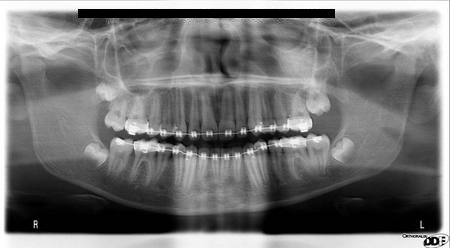
Figure 16: PROGRESS RECORDS 4-5-2010
I cannot provide accurate and comprehensive orthodontic diagnosis and treatment planning without a complete radiographic survey exam of our patients. Among the records provided us by our maxillofacial radiographic laboratory, my experience is the panoramic view is the most valuable. Even with the three dimensional aspects of data provided by the cone beam computerized tomography, I find the panoramic view essential in my daily orthodontic practice.
The Maxillofacial Radiographs Lab., that provides images to our orthodontic practice recently went through an upgrade to digital Panoramic and Lateral Cephalometric exposures. We receive the surveys by email usually the same day that the patient exposures are completed. This is a wonderful improvement for prompt service for our office and the patient. We in turn forward many of the views we receive to the family dentist and/or pedodontist by email as a routine office procedure. Once again our client families and dental practices see an attitude of providing a higher level of service thanks to the commitment to stay current made by our Maxillofacial Radiographic Laboratory.
The Maxillofacial Radiographs Lab., that provides images to our orthodontic practice recently went through an upgrade to digital Panoramic and Lateral Cephalometric exposures. We receive the surveys by email usually the same day that the patient exposures are completed. This is a wonderful improvement for prompt service for our office and the patient. We in turn forward many of the views we receive to the family dentist and/or pedodontist by email as a routine office procedure. Once again our client families and dental practices see an attitude of providing a higher level of service thanks to the commitment to stay current made by our Maxillofacial Radiographic Laboratory.