
Print Outs for dental digital imagesTarunjeet Pabla, BDS, MS Dip. ABOMR
Assistant Professor Division of Oral and Maxillofacial Radiology TUFTS University School of Dental Medicine Boston, MA From the Fall 2005 AADMRT Newsletter  Dr. Tarunjeet Pabla Dr. Tarunjeet Pabla
Digital radiography is increasingly being implemented in the field of dentistry. Images obtained from the digital dental radiographic systems are traditionally viewed on a CRT or a laptop display. These systems can be effectively used in a dental office setup with a personal computer. But in the case of an institutional setting where images are made in a radiology clinic and viewed in a different clinical area, a picture archival and communication system is required. Construction and implementation of such a system involves considerable investment of money and time. Until such a system is in place an alternative viewing method for dental digital images may be needed. Even if interpretation on a monitor makes film superfluous as a medium for diagnostics, it is unlikely that every dentist would have access to a digital viewing system. Some kind of hard copy for referral purposes will therefore, be needed.
Parallel to the increasing use of digital radiographic systems is a potential for utilization of computer peripheral devices. Ink jet printers have shown their potential for producing good quality photographs on glossy photographic paper. If the quality of images printed from such devices is sufficiently high then they can be used to print hard copies for general dental diagnostic tasks. We conducted a study to comparatively assess caries interpretation performance on various hard copy printing options with the soft copy (digital images). Digital images of 41 extracted posterior teeth were printed on glossy paper and film using HP DeskJet, and Codonics NP printers and compared to monitor viewed images. Ground truth was determined from histology. Performance of 9 observers and related factors were statistically analyzed. To obtain digital images, size 2 storage phosphor plates (Gendex® DenOptix® Imaging system) were exposed for 0.32 seconds and scanned in a DenOptix® scanner at 300dpi resolution. The images were displayed and sent to the printer through the DenOptix® system interface ( Dentsply Gendex VixWinTM 2000 software). |


|
|
Printers and Print media: The general purpose ink jet printers are relatively cheap and easy to use, hence the HP DeskJet 1220C printer (Hewlett-Packard Company, CA) was selected as one modality to print dental digital images.
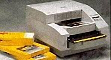
Another printer used was a Codonics NP 1660M Series Printer (Codonics, Inc. Middleburg Heights, OH) which represented the higher end of the cost spectrum and also represents dye-diffusion printing technology which is hailed as a superior technology when compared to ink-jet printing. 8.5 X 11 in. Kodak Distributed Medical Imaging Paper (Eastman Kodak Company, Rochester, NY) was used to print digital images. On subjective evaluation, the quality of printouts obtained on Kodak DMI paper were far more superior than other brand name glossy Photo papers available in the stores, hence it was decided to use Kodak paper. Chroma VistaTM Paper (8.5 X 10 in) and Direct VistaTM Film (8 x 10 in) (both recommended for the Codonics printer) were used for printing hard copies with the Codonics NP 1660M Series Printer Codonics NP 1660M Series Printer (Codonics, Inc.) Before the introduction of digital imaging systems, film mounted on a lighted view box was the most common method of diagnosing caries. Dentists are more used to looking at the films through transmitted light. Hence it was decided to print images on both transparent film as well as glossy paper. In order to obtain adequate print quality, subjective evaluation of the printouts was performed. Once a consensus was reached, those particular settings were selected on the printers to obtain hard copies of the test images. Print size: Printout size is an important issue. It not only affects the diagnostic capability but also the cost and time involved in printing the images. For example you can choose to print a 18- 20 images FMX on one sheet or you can choose to print one periapical size image on each sheet of paper.Print size is determined by the image system's display software, printer settings and print medium. A pilot study was conducted to determine a suitable print size for printing the images. Results of the pilot study showed that the bitewing (1.5x-magnified) format was significantly better than the Full mouth (unmagnified) format for proximal caries detection in printed images. The purpose of this study was to assess the performance of various print technologies in relation to digital images for proximal caries detection. The results of the study showed that the print outs were comparable to digital images in their diagnostic performance. A trend in improved caries detection performance was seen with Codonics printed hard copies when compared to the hard copies printed on inkjet printer. The Codonics printer prints on special paper with dye diffusion technology, which is considered a superior technology and is relatively expensive compared to the inkjet printing as provided HP printer. Because the results of our study showed no statistically significant difference in the diagnostic performance of the paper hard copies obtained from the two printers, its reasonable to say that for caries detection ink jet printers are up to the task. |
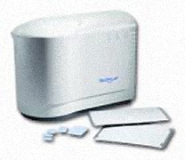
Gendex® DenOptix® Imaging system
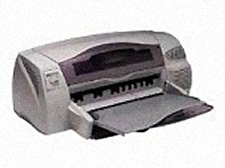
HP DeskJet 1220C printer (Hewlett-Packard Company, CA)
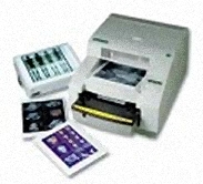
(Codonics, Inc. Middleburg Heights, OH)
Kodak 8650 and Medical Imaging Paper, Eastman Kodak Company, Rochester, NY)
|
Any new modality should fulfill at least the following three criteria: First, it should provide diagnostic information superior to or at least equal to the current system; second, it should be cost effective; and third it should be easy to operate. Hard copies obtained from the printers seemed to fulfill all these criteria. The initial cost involved with dye sublimation printer is high as compared to ink jet printers, but the cost of printed paper copy is less than $1 with the printers.
The hard copy printouts available currently have sufficient quality, even for diagnostic purposes. However, if digitization is to be meaningful and its potential fully exploited, the evaluation work should be based on monitor systems instead of hard copies. Paper might seem to be an inferior redundancy to film. But in fact, paper offers an economical and flexible medium that allows images to be used for tasks that previously have not included image display, such as patient report generation, student and employee instruction, and record keeping in research.
Our results suggest that caries diagnosing efficacy of hard copies was comparable to images displayed on CRT monitor. The option of hard copy certainly warrants exploration with other diagnostic tasks in dental radiography. This is especially true during periods of transition from analog to completely digital/electronic environments.