
SureSmile Applies CBCT to Custom Orthodontic TherapyEdward Y. Lin, DDS, MS* and Philip Getto**
*Orthodontic Specialists of Green Bay, Green Bay ,Wisconsin with contributions from Kevin J. Wilke, DDS, MS, Lwei M. Huang, DMD, MS, and Lee S Bialkowski, DDS **Chief Technology Officer OraMetrix, Richardson, Texas From the Winter 2010 AADMRT Newsletter
|


|
Although three-dimensional cone beam computed tomography (CBCT)has been available to the medical profession for more than 30 years, its high cost and radiation dosage, complexity, large footprint and resolution inadequacy for dental diagnostics has limited those in our profession from being able to capitalize on its use. With 3D CBCT accelerating to the forefront of diagnostic imaging, dental professionals can now avail themselves of relatively low radiation scans that create 360° virtual models. These models offer the clinical precision required for dental diagnostic analysis of the entire craniofacial structure with none of the distortion, magnification and superimposition problems of 2D radiographs (Figure 1). Just one scan provides images of the teeth, bone structure, TMJ, sinuses, nasal cavity, alveolar nerve canal and all related anatomy.
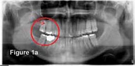
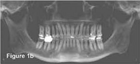
Figure 1: A comparison of a 2D panorex (a) with its elongated shapes and indistinct root structures with a 3D CBCT panorex (b) of the same patient clearly delineates space between the UR 1st and 2nd molars for placement of a temporary anchorage device - just one example of the benefit of CBCT-based images.
With CBCT, clinicians can view virtual patients from multiple perspectives in three planes of space, pinpointing the precise shape, location and morphology of unerupted teeth and dental roots within the bone. These 3D scans also offer measurements of bone density for accurate assesment of bone available for tooth movement. Pathologies (growths, bony dehiscences, fenestrations, etc.) are much more easily identified than through 2D X-rays (Figures 2a-b) as are skeletal asymmetries (Figures 3a-b).
With CBCT, clinicians can view virtual patients from multiple perspectives in three planes of space, pinpointing the precise shape, location and morphology of unerupted teeth and dental roots within the bone. These 3D scans also offer measurements of bone density for accurate assesment of bone available for tooth movement. Pathologies (growths, bony dehiscences, fenestrations, etc.) are much more easily identified than through 2D X-rays (Figures 2a-b) as are skeletal asymmetries (Figures 3a-b).

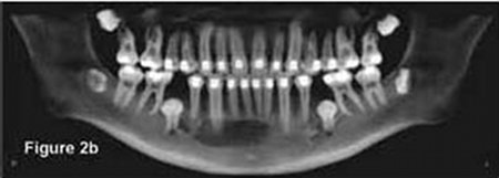
Figure 2: The 2D panorex of a patient taken in July, 2005 (a), does not make evident a traumatic bone cyst that was clearly revealed in the 3D CBCT panorex taken in January, 2007 (b).
The installed base of CBCT scanners in the U.S. is approximated at 2,500, which means that even at this early stage of commercial availability, at least 25% of orthodontists in the country have purchased a machine or have one available in their commmunity via an imaging center. The open environment and small footprint of CBCT scanning devices fit well in dental and orthodontic offices and radiation exposure has been reduced significantly from that of medical CT scanners. Cost of ownership is now reasonable vis-a-vis 2D radiographic machines and new management software has made their use much easier to master. These breakthroughs bode well for the complete migration from traditional radiography to 3D CBCT scanning in the not-too-distant future.
The installed base of CBCT scanners in the U.S. is approximated at 2,500, which means that even at this early stage of commercial availability, at least 25% of orthodontists in the country have purchased a machine or have one available in their commmunity via an imaging center. The open environment and small footprint of CBCT scanning devices fit well in dental and orthodontic offices and radiation exposure has been reduced significantly from that of medical CT scanners. Cost of ownership is now reasonable vis-a-vis 2D radiographic machines and new management software has made their use much easier to master. These breakthroughs bode well for the complete migration from traditional radiography to 3D CBCT scanning in the not-too-distant future.
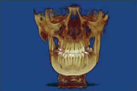
Figure 3: The 3D CBCT-based image of this patient's asymmetry (a) evidenced in her soft tissue (b) offers a superior diagnostic and treatment guide for the surgical recontouring of her inferior mandibular border.
SureSmie Extends CBCT with Therapeutic Application
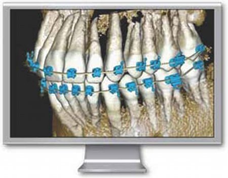
CBCT imaging, while immensely valuable for orthodontic, surgical and dental diagnostics, offers no direct means of using the data to change orthodontic treatment outcomes. For this capability, the SureSmile Digital Orthodontic System is a vital application that directly and positively affects such outcomes. The SureSmile system is comprised of unique treatment planning simulation software and a custom appliance design and manufacturing service. Having recognized SureSmile's potential to extend the capabilities of CBCT diagnostic technology into therapeutics, OraMetrix, the developers of SureSmile, had been monitoring the progress of its development and acceptance for some time. Four years ago, the company made the commitment to expend the considerable development resources required to integrate the two technologies, for the first time combining full-anatomy 3D diagnostics with custom therapeutics for orthodontic treatment (Figure 4) OraMetrix set for itself the standard of quality to which more than 17,000 SureSmile cases have already been successfully treated with high quality and improved efficiency: powerful simulation and evaluation decision support available at any time during treatment (even after brackets have been bonded) and accuracy to 50 microns. In May 2007, practices around the country began beta-testing the clinical application of the integration of CBCT into the SureSmile system. Today, OraMetrix offers these benefits to all SureSmile practitioners.
SureSmie Extends CBCT with Therapeutic Application
CBCT imaging, while immensely valuable for orthodontic, surgical and dental diagnostics, offers no direct means of using the data to change orthodontic treatment outcomes. For this capability, the SureSmile Digital Orthodontic System is a vital application that directly and positively affects such outcomes. The SureSmile system is comprised of unique treatment planning simulation software and a custom appliance design and manufacturing service. Having recognized SureSmile's potential to extend the capabilities of CBCT diagnostic technology into therapeutics, OraMetrix, the developers of SureSmile, had been monitoring the progress of its development and acceptance for some time. Four years ago, the company made the commitment to expend the considerable development resources required to integrate the two technologies, for the first time combining full-anatomy 3D diagnostics with custom therapeutics for orthodontic treatment (Figure 4) OraMetrix set for itself the standard of quality to which more than 17,000 SureSmile cases have already been successfully treated with high quality and improved efficiency: powerful simulation and evaluation decision support available at any time during treatment (even after brackets have been bonded) and accuracy to 50 microns. In May 2007, practices around the country began beta-testing the clinical application of the integration of CBCT into the SureSmile system. Today, OraMetrix offers these benefits to all SureSmile practitioners.
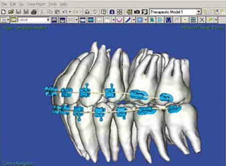
Figure 4: The first such interface of its kind, the accurate integration of 3D CBCT-based dental crown and root anatomy with the SureSmile therapeutic planning software brings distortion-free surface and subsurface morphology to SureSmile's 3D virtual setup simulations. This digital solution creates a precise virtual setup target for the fabrication of the SureSmile customized treatment appliance.
With this integrated solution, clinicians can now view the entire tooth anatomy (clinical crowns plus roots and other subsurface structures), manipulate teeth and jaws for simulating orthodontic treatment, and measure the changes any proposed treatment would make. From these virtual setups, individualized treatment is executed via SureSmile's robotically customized high-tech archwires. The integration of these two highly advanced technologies makes extensive clinical contributions to patient care.
With this integrated solution, clinicians can now view the entire tooth anatomy (clinical crowns plus roots and other subsurface structures), manipulate teeth and jaws for simulating orthodontic treatment, and measure the changes any proposed treatment would make. From these virtual setups, individualized treatment is executed via SureSmile's robotically customized high-tech archwires. The integration of these two highly advanced technologies makes extensive clinical contributions to patient care.
- There is a dramatic difference between simulating virtual treatment options with a complete understanding of the precise location, volume and orientation of a patient's actual root system versus using root structures based on anatomical norms based-estimations that are sometimes incorrect. A difference in direction or location of just one or two tooth roots can mean a significant difference in the way a clinician will treat the patient. This capability will be further advanced as SureSmile software incorpocates models of the bones from the CBCT scans.
- Since root resobrption of adjacent teeth is a major concern when planning the extrusion of unerupted and impacted teeth, CBCT-based treatment simulation with the SureSmile software is especially important in order to have as little impact as possible on (and further damaging) the roots of adjacent teeth (Figure 5) (For example, Walker, et al., reports research that found root resorption adjacent to impacted canines was 11.1% for central incisors and 66.7% for lateral incisors.1)
- Being able to capture the anatomy of teeth under soft tissue (whether wholly or partially erupted) also delineates what the size and orientation of the teeth will be when they erupt for accurate guidance and/or spatial planning (See Figure 6). Since SureSmile can now model unerupted teeth, it is possible to plan the desired final positions of these teeth precisely. Combined with the ability to manufacture appliances designed and refined virtually, the clinician is able to control their movement accurately after eruption and bonding.
- Planning for osseo-integrated implants requires optimal spacing and correct angulations of adjacent teeth, which CBCT/SureSmile integration makes feasible. At this point, CBCT images assist clinicians in assessing the quality of bone at the implant site.
- Clinicians can now easily build the trajectory of crown and root movement into the simulated treatment plan when using temporary anchorage devices.
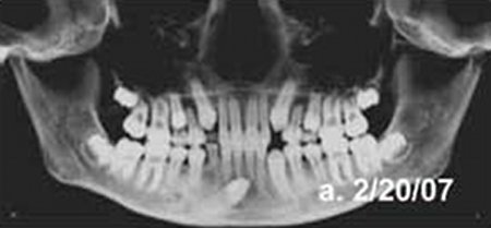

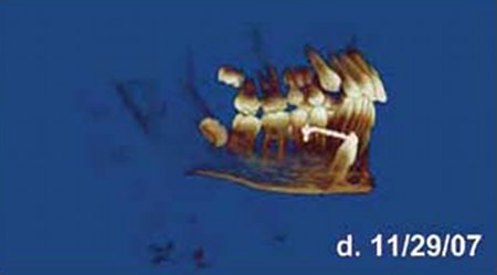
Figure 5: Having the roots as part of the SureSmile treatment planning simulation is especially important when planning the extrusion of unerupted or impacted teeth to have as little impact as possible on (and further damaging) the roots of adjacent teeth.
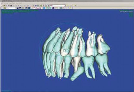
Figure 6: Since SureSmile can now model unerupted teeth, it is possible to plan the desired final positions of these teeth precisely. (The blue tint delineates the virtual diagnostic model; the white, the setup.)
Without full knowledge of root position and the bone available for tooth movement, clinicians must be somewhat conservative in crown movement. In conducting setup simulations, complete, anatomically correct data that includes bone and the volume, position and direction of the roots creates a highly accurate setup target. The more precise the target, the better the final root position. The better the final root position, the better the final crown position, the better the final occlusion and the quality, stability and esthetics of the result.
While offering accuracy comparable with the OraScanner (the scanning device SureSmile practitioners have used to date is the only solution that can be used in scme circumstances), CBCT images takes considerably less time to obtain. This time savings translates into significant additional production time for a busy orthodontic practice. Scans from Classic iCat® and Next Generation iCat® (Imaging Sciences, Hatfield, PA) take roughly 30 seconds (the time the patient must sit still for the imaging) and another 30 seconds for standard reconstruction, which translates the raw data into easily viewed images. Compared with the 20 minutes it takes to complete an OraScan of a patient, CBCT scanning also means a much more comfortable experience for the patient, an aspect of practice building that few orthodontisis take for granted.
Each of these benefits has the potential to enhance the reliability and quality of treatment and offer shorter treatment times. If, for example, an understanding of accurate root positions eliminates just one to two wire adjustments, that saving alone can drop treatment time by at least two months. With SureSmile treatment time averaging about 15 months, a two-month redudion in overall treatment time means a 13% pickup in production time. If, indeed, orthodontic treatment suffers from 30-40% relapse unless fixed retainers are worn for life, is it not reasonaable to assume that highly accurate setups, specifically in terms of root position in the bone, might conceivably impact these statistics in a positive direction?
What the Future Holds
The SureSmile software team is continuing development of the CBCT interface to extend its capabilities and remove current limitations. One can envision a future software release extracting maxillary and mandibular bone structures from the CBCT images for incorporation with the current SureSmile models of tooth and root anatomy. Such an enhancement will improve the confidence that target tooth placement is kept within the available bone. Information about the boundaries of supporting bone is particularly critical in cases where it is necessary to determine whether there is sufficient bone to resolve crowding without extraction or orthognathic surgery. Enhancements such as this will also be important in planning surgery and simulating its outcome. Being able to take measurements based on the relationship between the virtual setup and the achieved tocth position and supporting skeletal structure will yield the ultimate in orthodontic assesment systems.
The integration of CBCT images of dental root anatomy with SureSmile therapeutic planning software creates a precise virtual setup target for the fabrication of the SureSmile customized treatment appliance. The more precise the target the more refined the approach and the greater the efficiency and quality of treatment. This integration is a monumental achievement and is one for which the company and the clinicians who worked to make it happen are proud. What the long-term future holds is even more promising because this integration process is just the beginning of the journey. OraMetrix is committed to continuing its work to advance digital applications for orthodontic therapeutics, driving beyond the boundaries of these and related technologies for the benefit of its partner practices and their patients.
BIOGRAPHIES

Dr. Ed Lin and his partners in their three office practice, Orthodontic Specialists of Green Bay (OSGB), are strong proponents of incorporating advanced technologies for improving the efficiency and quality of patient care. They have an all-digital practice with digital records management and CBCT 3D imaging for diagnostics and treatment. The practice has been a fully committed SureSmile practice since 2005, and he has been beta testing the integration of CBCT scanning with SureSmile since early 2007. Dr. Lin earned his dental and orthodontic degrees at Northwestern University Dental School. He lectures nationally and volunteers at the Northeast Wisconsin Technical College Community Dental Clinic.
Philip Getto has spent more than 20 years in the industry developing commercial and technical software systems. He was recently senior vice president of MCAD product development and delivery at Paramentric Technology Corp, where he was responsible for all aspects and phases of software development and delivery effort for Pro/ENGINEER®, Pro/Intralink®, CADDS 5® Optegra® Pro/Desktop® and Medusa®, including product definition, program management, software development, testing, documentation, internationalization, integration and release management. While at PTC, he led the successful efforts to improve Pro/ENGINEER quality and usability. Mr. Getto has been manager of geometric modeling R&D for Rasna Corp., when it was acquired by Parametric. Mr. Getto received a bachelor of science and master of engineering degrees in computer and systems engineering at the Rensselaer Polytechnic Institute in Troy, N. Y.
REFERENCES
- Walker, L., Encisco, R., Mah, J.: Three dimensional localization of maxillary canines with cone-beam computed tomography, Am. J. Orthod. and Dentofacial Orthop., 2005: 128: pp. 418-423.