
Infection Control for dental radiographyCharles John Palenik, MS, PhD, MBA
Infection Control Research & Services Indiana University School of Dentistry Indianapolis, Indiana From the Fall 2004 AADMRT Newsletter 
During use, radiographic equipment can become contaminated use with patient blood and/or saliva if aseptic techniques are not practiced well. Dental health care personnel (DHCP) can also be at risk for occupational exposure. Oral microorganisms can remain viable on inert, radiographic equipment for more than 48 hours. Similar microbes have been shown to survive in x-ray developer/fixer for periods as long as two weeks.
Infection control practices for dental radiography are identical to those used in the operatory. They include standard precautions and are directed toward preventing disease transmission from patient to DHCP, from DHCP to patients, from patient to patient and from the practice to the surrounding locale. Aseptic procedures include the use of: 1) disposable and heat sterilizable x-ray accessories; 2) immersion of heat sensitive items in liquid chemical sterilant/high level disinfectants; 3) surface covers and intermediate level chemical disinfectants for clinical contact surfaces; 4) engineering and work practice controls; 5) personal protective devices (PPE) and 6) DHCP training. The greatest risk factor is when DHCP handle exposed radiographic films. Limiting the spread of body fluids present on such films is essential. Films used intraorally can become contaminated, handled and then transported throughout the practice environment. Also important is the covering or decontamination of soiled equipment. There are many environmental surfaces that could be touched. Examples include tube heads, extension cones, control panels, exposure buttons, chair controls, film processors, surfaces in darkrooms and any other area touched by contaminated films, gloved hands or equipment used orally. |


|
Infection control procedures for dental radiography can be divided into segments - activities before taking radiographs, activities performed while taking a radiograph, things to do after radiographs have been taken and finally, tasks associated with film processing. Suggested activities that follow apply to films held within barrier protective pouches. Some films come pouched by the manufacturer or pouches can be purchased separately.
Advice on using x-ray films without barrier pouches, taking panoramic/cephalometric x-rays, using a daylight loader for processing and the use of digital radiography sensors will then be given.
Advice on using x-ray films without barrier pouches, taking panoramic/cephalometric x-rays, using a daylight loader for processing and the use of digital radiography sensors will then be given.
Using Intraoral Films Held within Barrier Pouches
Preparing to Take Dental Radiographs
It is important to prepare the area prior to the seating the patient. Preparation increases the chances of success. Items used in the mouth should either be single use, disposable or sterilized by heat.
It is important to prepare the area prior to the seating the patient. Preparation increases the chances of success. Items used in the mouth should either be single use, disposable or sterilized by heat.
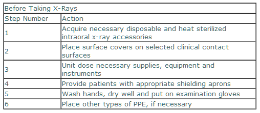
Covers generally involve plastic sheets, tubes or pieces with adhesive edges. Surfaces best covered include chair headrest and control adjustments, exposure buttons, control panels and x-ray tube heads and yokes.
Unit dosing: 1) reduces the need to leave the immediate area to search of needed items; 2) decreases the chances of DHCP contaminating large boxes or storage containers materials and 3) reduces the chances of cross-contamination of environmental surfaces. Things best unit dosed include paper towels, mouth props, film holders (e.g., cups), gloves, surface covers, film packs and cotton rolls.
Barriers: 1) protect films from direct contamination; 2) reduce the time needed for preparation and processing by eliminating the need for disinfection in the darkroom and 3) decreases the necessity of wearing additional pairs of gloves. Film barriers are the method of choice especially when using daylight loaders. Their use eliminates almost all chances of equipment contamination.
After the patient has been seated, hands can be washed and dried and gloves placed. Then, heat sterilized x-ray holding/positioning devices can be removed from their packages and assembled in view of the patient.
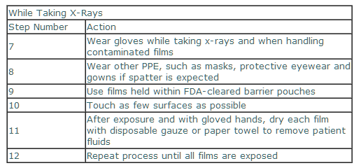
Taking Dental Radiographs
Gloves should be worn when taking radiographs and handling contaminated film packets. Other PPE should be used when spattering of patient body fluids is likely. Ideally, only heat tolerant intraoral x-ray accessories (e.g., film holders and positioning devices) should be used. Some items, such as mouth props can either be sterilized or be single use disposable.
Unit dosing: 1) reduces the need to leave the immediate area to search of needed items; 2) decreases the chances of DHCP contaminating large boxes or storage containers materials and 3) reduces the chances of cross-contamination of environmental surfaces. Things best unit dosed include paper towels, mouth props, film holders (e.g., cups), gloves, surface covers, film packs and cotton rolls.
Barriers: 1) protect films from direct contamination; 2) reduce the time needed for preparation and processing by eliminating the need for disinfection in the darkroom and 3) decreases the necessity of wearing additional pairs of gloves. Film barriers are the method of choice especially when using daylight loaders. Their use eliminates almost all chances of equipment contamination.
After the patient has been seated, hands can be washed and dried and gloves placed. Then, heat sterilized x-ray holding/positioning devices can be removed from their packages and assembled in view of the patient.
Taking Dental Radiographs
Gloves should be worn when taking radiographs and handling contaminated film packets. Other PPE should be used when spattering of patient body fluids is likely. Ideally, only heat tolerant intraoral x-ray accessories (e.g., film holders and positioning devices) should be used. Some items, such as mouth props can either be sterilized or be single use disposable.
After Taking Dental Radiographs
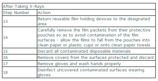
Care must be taken when opening protective film barriers. Unsoiled films are the desired goal. Handling such items without gloves makes processing much easier in the darkroom and daylight loader.
Care must be taken when opening protective film barriers. Unsoiled films are the desired goal. Handling such items without gloves makes processing much easier in the darkroom and daylight loader.
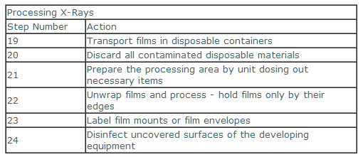
Developing Dental Radiographs
Once in the developing area, open film packets with clean, ungloved hands. Holding the tab, films can go into clean plastic cups or onto paper towels. Holding films by their edges, insert them into the processor. Just to be safe, it is best to either cover or regularly clean and disinfect the surfaces of developing equipment.
Once in the developing area, open film packets with clean, ungloved hands. Holding the tab, films can go into clean plastic cups or onto paper towels. Holding films by their edges, insert them into the processor. Just to be safe, it is best to either cover or regularly clean and disinfect the surfaces of developing equipment.
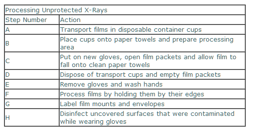
Using Intraoral Films Not Held within Barrier Pouches
It is important when handling and processing films not held in protective barriers that they be well wiped off chairside. It is very important to either cover or clean and disinfect any surface that may become contaminated.
It is important when handling and processing films not held in protective barriers that they be well wiped off chairside. It is very important to either cover or clean and disinfect any surface that may become contaminated.
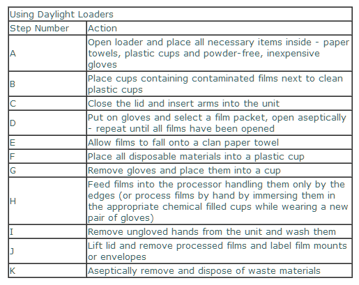
Processing X-Ray Films in a Daylight Loader
Daylight loaders have cloth or rubber sleeves, cuffs or flaps that allow for the passage of materials/arms without permitting light to enter. Such equipment is difficult to impossible to clean or disinfect. It is imperative that an effective, yet efficient protocol for aseptic use be established and rigorously maintained. After reviewing the following steps, it should be obvious how useful barrier protected films are.
Daylight loaders have cloth or rubber sleeves, cuffs or flaps that allow for the passage of materials/arms without permitting light to enter. Such equipment is difficult to impossible to clean or disinfect. It is imperative that an effective, yet efficient protocol for aseptic use be established and rigorously maintained. After reviewing the following steps, it should be obvious how useful barrier protected films are.
Digital X-Rays and X-Rays
Digital radiographic sensors and other high technology instruments, such as intraoral camera, electronic periodontal probes, colossal analyzers and lasers come into contact with mucus membranes. Such devices are considered to be semicritical items. Ideally, they should be cleaned and heat sterilized or high level disinfected between patients. However, many cannot be heat sterilized, nor undergo high level disinfection.
Semicritical items that cannot be reprocessed by heat sterilization or high level disinfection should, at a minimum be barrier protected by using a FDA cleared barrier to reduce gross contamination. Covers are not always totally protective. So, after removing the barriers, devices should be cleaned and disinfected using an intermediate level disinfectant (tuberculocidal) after each patient. Manufacturers should be consulted as to sterilization and disinfection processes, as well as the types of covers to be used.
Summary
Infection control for dental radiography employs the same materials, processes and techniques used in the operatory. Unless proper procedures are established and followed, there is a definite potential for cross-contamination to clinical area surfaces and DHCP. In general, the aseptic practices used are relatively simple and inexpensive; yet require complete application in every situation.
Recently, the Organization for Safety & Asepsis Procedures (OSAP) produced a workbook (From Policy to Practice: OSAP?s Guide to the Guidelines), which covers the materials presented in the new CDC infection control recommendations. Seven pages of the workbook specifically address radiographic infection control. More information can be obtained on the OSAP websitewww.osap.org.
Charles John Palenik, MS, PhD, MBA
Infection Control Research & Services
Indiana University School of Dentistry
1121 West Michigan Street
Indianapolis, Indiana USA
voice - 317.274.5427
fax - 317.274.5428