
Radiographic Exam for the Pregnant Dental PatientDr. Dania Tamimi
Diplomate, American Board of Oral and Maxillofacial Radiology Private Practice Orlando, FL From the Spring 2008 AADMRT Newsletter  Dr. Dania Tamimi Dr. Dania Tamimi
Dental radiography is a controversial area in the management of the pregnant patient. In pamphlets widely supported by most dental professional organizations, no alteration of recommendation was given for prescribing radiographs to a pregnant patient, as the amount of radiation given during standard dental radiographic examination is so trivial that it could not cause gross anatomic malformations in the developing fetus.
The American Dental Association (ADA) recommends every precaution should be taken to minimize radiation exposure to the pregnant patient. The National Commission of Radiation Protection (NCRP) reports that production of congenital defects is negligible from fetal exposures of 50 mSv. Yet, in a questionnaire study of 552 dentists, almost all saw pregnancy as contradictory for bitewing radiographs, thus showing the need for clinician education using evidence-based dentistry. The estimated fetal doses from typical radiographic examinations lend support to the conclusion that fetal risks are minimal and, therefore, radiologic examinations that may provide significant diagnostic information should not be withheld from pregnant women. This is the position advocated by the International Commission on Radiation protection, American College of Radiology, and American College of Obstetrics and Gynecology. Needless to say, any potentially harmful factors that may affect the unborn child should be avoided, especially during the first trimester, and the As Low As Reasonably Achievable (ALARA) concept should be used as with all other patients. Effects of radiation exposure during prenatal development: To understand the effects of ionizing radiation on the unborn child, it is important to understand the units of radiation measurement:
|


|
The amount of radiation in standard dental radiographic examination as compared to natural radiation:
The adverse effects that may occur to the fetus of an expectant mother irradiated with high doses of radiation depend upon the fetal age and the dose of the ionizing radiation given. The most vulnerable time is during the first trimester:
Precautions to be taken when subjecting a pregnant patient to radiation:
As can be noted, most of these recommendations are an application of the ALARA rule and are the same precautions that should be taken for any patient imaged radiographically. The first two precautions are specific to the pregnant or possibly pregnant patient to avoid exposure of the abdomen with even the most negligible amount of radiation. Radiologic examinations should be performed only when necessary, and - as with any drug or intervention in pregnancy - the dose used for the examination should be kept as low as reasonably achievable.
- The universe and our industrialized world give off a certain amount of radiation per day (the average effective dose for a member of the US population is mSv a year).
- If the amount of radiation in standard plain film radiographic examinations was to be compared to the radiation that an individual receives from natural and artificial sources every day, it would negligible.
The adverse effects that may occur to the fetus of an expectant mother irradiated with high doses of radiation depend upon the fetal age and the dose of the ionizing radiation given. The most vulnerable time is during the first trimester:
- During implantation of the fertilized ovum: If a dose of 0.2 Gy or higher is given, death of the embryo may occur.
- During the first two months (organogenesis): a dosage of 0.2 Gy may cause macroscopic anatomical malformation.
- Between 8-15 weeks (fetogenesis): this is a period of high radiosensitivity for the developing central nervous system. Research has shown that a dosage of 1 Gy during this period caused mental retardation of 50% of fetuses studied.
Precautions to be taken when subjecting a pregnant patient to radiation:
- Observation of the "Ten-Day Rule": Any woman of childbearing age to be subjected to diagnostic x-ray examination that may reach the abdominal or pelvic areas should be exposed only during the first ten days after menstruation.
- The maxillary occlusal view or any other view that requires the x-ray beam passing down into the abdominal area should be avoided if proper shielding cannot be provided.
- Elective radiographs should be avoided.
- For emergency treatment, necessary radiographs should be limited to the areas in question.
- Try to minimize errors and retakes.
- Use of E-speed or Ekta plus speed film if using analog radiography: the faster the film, the less radiation exposure to the patient.
- Switching to digital radiography (decreases the does about 47% for full mouth series, and about 17% for panoramic).
- Use of thyroid shields.
- Use of lead aprons to cover the abdominal and pelvic areas.
- Maintain high beam energy to deliver a high quality diagnostic x-ray beam in the shortest possible time.
- Use of a long rectangular cone for collimation.
- Lower mAs setting on CBCT to decrease dose.
- Limitation of the field of view (FOV) on CBCT as indicated to give the necessary information for treatment planning without exposing unnecessary structures (example: narrowing the FOV for the open scan for TMJ to include just the TMJs structures, or limited maxillary or mandibular views for implant treatment planning).
As can be noted, most of these recommendations are an application of the ALARA rule and are the same precautions that should be taken for any patient imaged radiographically. The first two precautions are specific to the pregnant or possibly pregnant patient to avoid exposure of the abdomen with even the most negligible amount of radiation. Radiologic examinations should be performed only when necessary, and - as with any drug or intervention in pregnancy - the dose used for the examination should be kept as low as reasonably achievable.
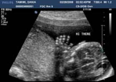
Dr. Tamimi is expecting her second child in July 2008, this is a sonogram of Dr. Tamimi's baby girl.
REFERENCES
- White S, Pharoah M: Oral radiology principles and practice, 5th edition. Mosby, Inc, St. Louis Missouri.
- Hall E: Radiobiology for the Radiologist, 5th ed. 2000, Lippencott Williams & Wilkins, Philadelphia, PA.
- McCollough C et al: Radiographics, Radiation Exposure and Pregnancy: When Should We Be Concerned? Volume 27, #4, July-Aug 2007: 909-918
- Rushton VE, Horne K, Worthington HV. Factors influencing the frequency of bitewing radiography in general dental practice. Comm Dent Oral Epidemiol 1996; 24:272-6
- Kan MWK, Leung LHT, Wong W, Lam N:Radiation Dose from Cone Beam Computed Tomography for Image-Guided Radiation Therapy. Int J Radiat Oncol Biol Phys. 2007 Oct 31; : 17980510 (P,S,E,B,D)
- Lambrecht JT, J. Roth J, Kiefer HP: Dose exposition from intra- and extraoral dental radiography CARS 2004 - Computer Assisted Radiology and Surgery. Proceedings of the 18th International Congress and Exhibition, International congress series, Volume 1268, June 2004, Pages 1147-1151